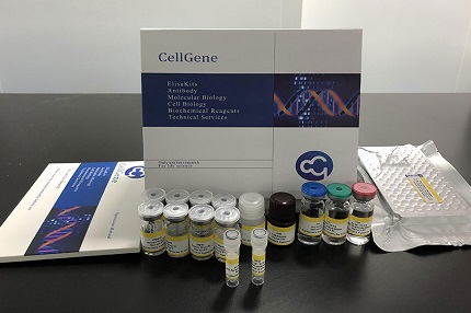
E03A0669 Mouse 26S Proteasome ELISA kit
The Mouse 26S Proteasome ELISA kit can be used to identify samples from the mouse species. 26S Proteasome can also be called 26S proteasome, 26S PSM.


E03A0669 Mouse 26S Proteasome ELISA kit
The Mouse 26S Proteasome ELISA kit can be used to identify samples from the mouse species. 26S Proteasome can also be called 26S proteasome, 26S PSM.
Product Information | |
Cat. No. | E03A0669 |
Product Name | Mouse 26S Proteasome ELISA kit |
Species | Mouse |
Product Size | 48 Tests / 96 Tests |
Concentration | 5.0-100ng/ml |
Sensitivity | 1.0 ng/mL |
Principal | Competitive ELISA |
Sample Volume | 100 ul |
Sample Type | Serum, plasma, cell culture supernatants, body fluid and tissue homogenate |
Assay Time | 90 minutes |
Platform | Microplate Reader |
Conjugate | HRP |
Detection Method | Colorimetric |
Storage | 2-8°C |
Kit Components | ||
MATERIALS | SPECIFICATION | QUANTITY |
MICROTITER PLATE | 96 wells | stripwell |
ENZYME CONJUGATE | 6.0 mL | 1 vial |
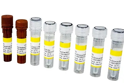
STANDARD A (0.5mL) | 0 ng/mL | 1 vial |
STANDARD B (0.5mL) | 5.0 ng/mL | 1 vial |
STANDARD C (0.5mL) | 10 ng/mL | 1 vial |
STANDARD D (0.5mL) | 25 ng/mL | 1 vial |
STANDARD E (0.5mL) | 50 ng/mL | 1 vial |
STANDARD F (0.5mL) | 100 ng/mL | 1 vial |
SUBSTRATE A | 6 mL | 1 vial |
SUBSTRATE B | 6 mL | 1 vial |
STOP SOLUTION | 6 mL | 1 vial |
WASH SOLUTION (100 x) | 10 mL | 1 vial |
BALANCE SOLUTION | 3 mL | 1 vial |
Principle of the Assay |
26S PSM ELISA kit applies the competitive enzyme immunoassay technique utilizing an anti-26S PSM antibody and a 26S PSM-HRP conjugate. The assay sample and buffer are incubated together with 26S PSM-HRP conjugate in pre-coated plate for one hour. After the incubation period, the wells are decanted and washed five times. The wells are then incubated with a substrate for HRP enzyme. The product of the enzyme-substrate reaction forms a blue colored complex. Finally, a stop solution is added to stop the reaction, which will then turn the solution yellow. The intensity of color is measured spectrophotometrically at 450nm in a microplate reader. The intensity of the color is inversely proportional to the 26S PSM concentration since 26S PSM from samples and 26S PSM-HRP conjugate compete for the anti-26S PSM antibody binding site. Since the number of sites is limited, as more sites are occupied by 26S PSM from the sample, fewer sites are left to bind 26S PSM-HRP conjugate. A standard curve is plotted relating the intensity of the color (O.D.) to the concentration of standards. The 26S PSM concentration in each sample is interpolated from this standard curve. |
Coefficient of Variance | Intra Variation% <10% | |
Inter Variation% <12% | ||
Recovery | 95-102% | |
Linearity | Diluent Ratio | Range % |
1:2 | 93-105 | |
1:4 | 88-106 | |
1:8 | 86-108 | |
Specificity/Cross-reactivity | No significant cross-reactivity or interference between 26S PSM and analogues was observed. | |


E03A0669 has been referenced in the below publications:
Coordinated regulation of protein synthesis and degradation by mTORC1.
Related Bluegene Biotech Products
BlueGene Biotech News


