E02S0011 Rat Stem Cell Factor ELISA kit
The Rat Stem Cell Factor ELISA kit can be used to identify samples from the rat species. Stem Cell Factor can also be called KITLG, FPH2, FPHH, KL-1, Kitl, MGF, SCF, SF, SHEP7, DCUA, KIT ligand, DFNA69, SLF.


E02S0011 Rat Stem Cell Factor ELISA kit
The Rat Stem Cell Factor ELISA kit can be used to identify samples from the rat species. Stem Cell Factor can also be called KITLG, FPH2, FPHH, KL-1, Kitl, MGF, SCF, SF, SHEP7, DCUA, KIT ligand, DFNA69, SLF.
Product Information | |
Cat. No. | E02S0011 |
Product Name | Rat Stem Cell Factor ELISA kit |
Species | Rat |
Product Size | 48 Tests / 96 Tests |
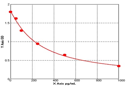
Concentration | 50-1000 pg/mL |
Sensitivity | 1.0 pg/ml |
Principal | Sandwich ELISA |
Sample Volume | 50 ul |
Sample Type | Serum, plasma, cell culture supernatants, body fluid and tissue homogenate |
Assay Time | 90 minutes |
Platform | Microplate Reader |
Conjugate | HRP |
Detection Method | Colorimetric |
Storage | 2-8°C |
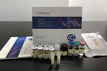
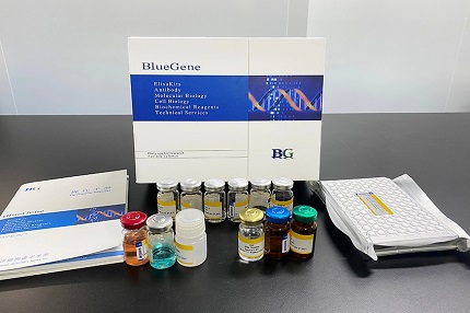
Kit Components | ||
MATERIALS | SPECIFICATION | QUANTITY |
MICROTITER PLATE | 96 wells | stripwell |
ENZYME CONJUGATE | 10 mL | 1 vial |
STANDARD A (0.5mL) | 0 pg/ml | 1 vial |
STANDARD B (0.5mL) | 50 pg/ml | 1 vial |
STANDARD C (0.5mL) | 100 pg/ml | 1 vial |
STANDARD D (0.5mL) | 250 pg/ml | 1 vial |
STANDARD E (0.5mL) | 500 pg/ml | 1 vial |
STANDARD F (0.5mL) | 1000 pg/ml | 1 vial |
SUBSTRATE A | 6 mL | 1 vial |
SUBSTRATE B | 6 mL | 1 vial |
STOP SOLUTION | 6 mL | 1 vial |
WASH SOLUTION (100 x) | 10 mL | 1 vial |
BALANCE SOLUTION | 3 mL | 1 vial |
Principle of the Assay |
SCF ELISA kit applies the quantitative sandwich enzyme immunoassay technique. The microtiter plate has been pre-coated with a monoclonal antibody specific for SCF. Standards or samples are then added to the microtiter plate wells and SCF if present, will bind to the antibody pre-coated wells. In order to quantitatively determine the amount of SCF present in the sample, a standardized preparation of horseradish peroxidase (HRP)-conjugated polyclonal antibody, specific for SCF are added to each well to “sandwich” the SCF immobilized on the plate. The microtiter plate undergoes incubation, and then the wells are thoroughly washed to remove all unbound components. Next, substrate solutions are added to each well. The enzyme (HRP) and substrate are allowed to react over a short incubation period. Only those wells that contain SCF and enzyme-conjugated antibody will exhibit a change in color. The enzyme-substrate reaction is terminated by addition of a sulphuric acid solution and the color change is measured spectrophotometrically at a wavelength of 450 nm. A standard curve is plotted relating the intensity of the color (O.D.) to the concentration of standards. The SCF concentration in each sample is interpolated from this standard curve. |
Coefficient of Variance | Intra Variation% <10% | |
Inter Variation% <12% | ||
Recovery | 95-102% | |
Linearity | Diluent Ratio | Range % |
1:2 | 93-105 | |
1:4 | 88-106 | |
1:8 | 86-108 | |
Specificity/Cross-reactivity | No significant cross-reactivity or interference between SCF and analogues was observed. | |


E02S0011 has been referenced in the below publications:
Preparation of FSH and SCF microspheres and sustained released study in vitro and in vivo.
Study on effects of the spermatogenesis of NOA model treated with testicular sustained-release FSH and SCF.
Preparation of the Sustain Release Performance of FSH and SCF Microspheres.
Related Bluegene Biotech Products


